WoRMS taxon details
Abyssocladia falkor Ekins & Hooper, 2023
1666703 (urn:lsid:marinespecies.org:taxname:1666703)
accepted
Species
marine, fresh, terrestrial
recent only
Ekins, M.; Hooper, J.N.A. (2023). New carnivorous sponges from the Great Barrier Reef, Queensland, Australia collected by ROV from the RV FALKOR. <em>Zootaxa.</em> 5293 (3): 435-471., available online at https://doi.org/10.11646/zootaxa.5293.3.2
page(s): 440-442 [details] Available for editors [request]
page(s): 440-442 [details] Available for editors
Holotype QM G339306, geounit Coral Sea
Holotype QM G339306, geounit Coral Sea [details]
Description Growth form: The holotype consists of a pedunculated sponge, with a short stem and a disc shaped body with filaments...
Distribution This species is currently known from the Coral Sea in Queensland, Australia, bathyal depth.
Etymology Falkor, a noun in apposition, named in honour of the Schmidt Ocean Institutes research vessel the RV Falkor.
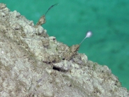
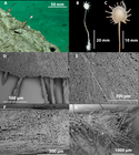
Description Growth form: The holotype consists of a pedunculated sponge, with a short stem and a disc shaped body with filaments radiating out in a single plane from the disc margin. (Figs. 4 A–C). The paratype has the entire stem and roots as well, which were lost from the holotype during collection. The holotype has a body diameter of 7.8 mm, and 1.3 mm thick. The filaments are 2.3 mm long and 0.1 mm in width. The paratype has a body with a 6.7 mm diameter and 1.6 mm width, the stem and root are 40 mm in length, the filaments are up to 4.0 mm in length.
Colour: Cream in situ, on deck and in ethanol.
Ectosomal skeleton: Thin membranous layer of the filaments is encrusted with small sigmancistras and abyssochelae. The sponge body and stem are encrusted with the predominately spherical microstrongyles, but also include the abyssochelae. Immediately under the encrusting layer are the sinuous styles. The ectosomal skeleton of the roots is a thin membranous layer with occasional abyssochelae, sigmas and sigmancistras.
Endosomal skeleton: The axis of the stem consists of longitudinally arranged mycalostyles, styles and sinuous styles (Figs. 5 D, F, J). The main body consists of the radiating bundles of subtylostyles that form the filaments (Figs. 5 H, I). Within the body between the bundles, the styles form a disorganised almost cross hatching skeleton. The roots are composed of the large mycalostyles, styles, sinuous styles, occasional subtylostyles and rarely strongyles (Fig. 4 G). The filaments axis is composed of subtylostyles only (Fig. 4 D).
Megascleres: Mycalostyles are long and generally straight, sometimes with a slight curve, and have a rounded point (Figs. 5 D, E). Styles usually straight, sometimes with a slight curve, thickest in the centre also with a blunt point, sometimes with a very faint tyle swelling (Figs. 5 F, G). They are separated from the mycalostyles often by their location in the body but could well be a continuum of a single category of megasclere. The sinuous styles occur around the mycalostyles that make up the stem, under the dermal microstrongyles (Figs. 5 J, K). Subtylostyles are long and straight, with a sharp tip and compose the filaments (Figs. 5 H, I). There are also very rare strongyles present from the roots (Figs. 5 L, M).
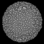
Microscleres: Abyssochelae isochelae with curved shaft, and opposing alae touching (Fig. 5 A). There are small sigmas and small sigmancistras that usually occur on the exterior surfaces (Figs. 5 C, N, respectively). There are also long thin sigmas that only occur on the sponge body, these were rare and only occurred on the paratype and so may not be native (Fig. 5 B). The predominately spherical to subspherical shaped microstrongyles are microspined (Figs. 4 E, F; 5 O). However, they are also dumbbell-shaped and rarely dildo—shaped (Fig. 5 P). The microstrongyles have either a rounded end (tylomicrostrongyle) or a sharp point (tylomicrostyles). [details]
Distribution This species is currently known from the Coral Sea in Queensland, Australia, bathyal depth.
Distribution This species is currently known from the Coral Sea in Queensland, Australia, bathyal depth. [details]
Etymology Falkor, a noun in apposition, named in honour of the Schmidt Ocean Institutes research vessel the RV Falkor.
Etymology Falkor, a noun in apposition, named in honour of the Schmidt Ocean Institutes research vessel the RV Falkor. [details]
de Voogd, N.J.; Alvarez, B.; Boury-Esnault, N.; Cárdenas, P.; Díaz, M.-C.; Dohrmann, M.; Downey, R.; Goodwin, C.; Hajdu, E.; Hooper, J.N.A.; Kelly, M.; Klautau, M.; Lim, S.C.; Manconi, R.; Morrow, C.; Pinheiro, U.; Pisera, A.B.; Ríos, P.; Rützler, K.; Schönberg, C.; Turner, T.; Vacelet, J.; van Soest, R.W.M.; Xavier, J. (2024). World Porifera Database. Abyssocladia falkor Ekins & Hooper, 2023. Accessed through: World Register of Marine Species at: https://www.marinespecies.org/aphia.php?p=taxdetails&id=1666703 on 2024-11-22
Date
action
by
![]() The webpage text is licensed under a Creative Commons Attribution 4.0 License
The webpage text is licensed under a Creative Commons Attribution 4.0 License
original description
Ekins, M.; Hooper, J.N.A. (2023). New carnivorous sponges from the Great Barrier Reef, Queensland, Australia collected by ROV from the RV FALKOR. <em>Zootaxa.</em> 5293 (3): 435-471., available online at https://doi.org/10.11646/zootaxa.5293.3.2
page(s): 440-442 [details] Available for editors [request]
page(s): 440-442 [details] Available for editors
 Present
  Present in aphia/obis/gbif/idigbio
  Inaccurate
  Introduced: alien
  Containing type locality
Holotype QM G339306, geounit Coral Sea [details]
From editor or global species database
Description Growth form: The holotype consists of a pedunculated sponge, with a short stem and a disc shaped body with filaments radiating out in a single plane from the disc margin. (Figs. 4 A–C). The paratype has the entire stem and roots as well, which were lost from the holotype during collection. The holotype has a body diameter of 7.8 mm, and 1.3 mm thick. The filaments are 2.3 mm long and 0.1 mm in width. The paratype has a body with a 6.7 mm diameter and 1.6 mm width, the stem and root are 40 mm in length, the filaments are up to 4.0 mm in length.Colour: Cream in situ, on deck and in ethanol.
Ectosomal skeleton: Thin membranous layer of the filaments is encrusted with small sigmancistras and abyssochelae. The sponge body and stem are encrusted with the predominately spherical microstrongyles, but also include the abyssochelae. Immediately under the encrusting layer are the sinuous styles. The ectosomal skeleton of the roots is a thin membranous layer with occasional abyssochelae, sigmas and sigmancistras.
Endosomal skeleton: The axis of the stem consists of longitudinally arranged mycalostyles, styles and sinuous styles (Figs. 5 D, F, J). The main body consists of the radiating bundles of subtylostyles that form the filaments (Figs. 5 H, I). Within the body between the bundles, the styles form a disorganised almost cross hatching skeleton. The roots are composed of the large mycalostyles, styles, sinuous styles, occasional subtylostyles and rarely strongyles (Fig. 4 G). The filaments axis is composed of subtylostyles only (Fig. 4 D).
Megascleres: Mycalostyles are long and generally straight, sometimes with a slight curve, and have a rounded point (Figs. 5 D, E). Styles usually straight, sometimes with a slight curve, thickest in the centre also with a blunt point, sometimes with a very faint tyle swelling (Figs. 5 F, G). They are separated from the mycalostyles often by their location in the body but could well be a continuum of a single category of megasclere. The sinuous styles occur around the mycalostyles that make up the stem, under the dermal microstrongyles (Figs. 5 J, K). Subtylostyles are long and straight, with a sharp tip and compose the filaments (Figs. 5 H, I). There are also very rare strongyles present from the roots (Figs. 5 L, M).
Microscleres: Abyssochelae isochelae with curved shaft, and opposing alae touching (Fig. 5 A). There are small sigmas and small sigmancistras that usually occur on the exterior surfaces (Figs. 5 C, N, respectively). There are also long thin sigmas that only occur on the sponge body, these were rare and only occurred on the paratype and so may not be native (Fig. 5 B). The predominately spherical to subspherical shaped microstrongyles are microspined (Figs. 4 E, F; 5 O). However, they are also dumbbell-shaped and rarely dildo—shaped (Fig. 5 P). The microstrongyles have either a rounded end (tylomicrostrongyle) or a sharp point (tylomicrostyles). [details]
Distribution This species is currently known from the Coral Sea in Queensland, Australia, bathyal depth. [details]
Etymology Falkor, a noun in apposition, named in honour of the Schmidt Ocean Institutes research vessel the RV Falkor. [details]
Remark This species is most closely related to the Japanese species A. natsushimae Ise & Vacelet, 2010. However, this new species has a much smaller and circular body size. The new species also lacks the two size classes of abyssochelae, has subtylostyles, and has only one category of sigmancistras and two categories of sigmas. The microstrongyles of this new species are predominately spherical and lacks the long microstrongyles of A. natsushimae. This species is different from the diminutive species A. bruuni Lévi, 1964, which has the very large abyssochelae. Abyssocladia bruuni was also recorded by Koltun (1970), but with two size classes of abyssochelae. Abyssocladia bruuni also lack microstrongyles. This new species also has similar abyssochelae and a rough morphological similarity to A. annae Ekins et al., 2020a and another closely related species A. fryerae Hestetun et al., 2019, both of which have this ridged abyssochelae as well as subtylostyles and sigmancistras. [details]


