Integrated Marine Information System (IMIS)
Persons | Institutes | Publications | Projects | DatasetsAphanostoma pisae
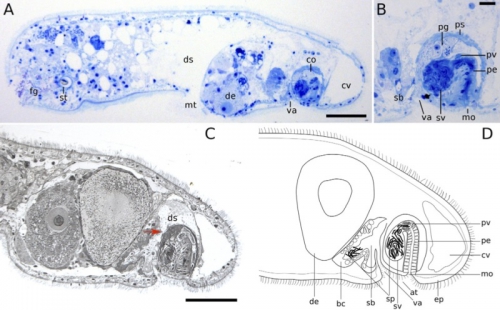
Description Sagittal sections of mature animals, anterior to the left, dorsal up. (?B) Stained after Richardson et al. (1960). (C) Stained after Heidenhain (Romeis, 1968). Red arrow marks the opening of the bursa to the digestive syncytium. (D) Schematic drawing, based on sections of several individuals. at atrium, bc bursal cap, co copulatory apparatus, cv chordoid vacuole, de developing egg, ds digestive syncytium, ep ciliated epidermis, fg frontal glands, mo male genital opening, mt mouth, pe penis, pg prostatic glands, ps penis sheath, pv prostatic vesicle, sb seminal bursa, sp sperm, st statocyst, sv seminal vesicle, va vagina. Scale bars: 50 ?m in (A, C), 10 ?m in (B)
JPG file - 129.63 kB - 800 x 496 pixels
Extra information
FileName: 124569.jpg
FileDateTime: 1427960116
FileSize: 129626
FileType: 2
MimeType: image/jpeg
SectionsFound: ANY_TAG, IFD0, EXIF
COMPUTED.html: width="800" height="496"
COMPUTED.Height: 496
COMPUTED.Width: 800
COMPUTED.IsColor: 1
COMPUTED.ByteOrderMotorola: 1
XResolution: 150/1
YResolution: 150/1
ResolutionUnit: 2
Exif_IFD_Pointer: 78
ExifImageWidth: 961
ExifImageLength: 596
added on 2017-04-071 704 views
FileName: 124569.jpg
FileDateTime: 1427960116
FileSize: 129626
FileType: 2
MimeType: image/jpeg
SectionsFound: ANY_TAG, IFD0, EXIF
COMPUTED.html: width="800" height="496"
COMPUTED.Height: 496
COMPUTED.Width: 800
COMPUTED.IsColor: 1
COMPUTED.ByteOrderMotorola: 1
XResolution: 150/1
YResolution: 150/1
ResolutionUnit: 2
Exif_IFD_Pointer: 78
ExifImageWidth: 961
ExifImageLength: 596
Click here to return to the thumbnails overview
