Integrated Marine Information System (IMIS)
Persons | Institutes | Publications | Projects | DatasetsRamisyllis kingghidorahi Aguado, Ponz-Segrelles, Glasby, Ribeiro, Jimi & Miura, In Aguado, Ponz-Segrelles, Glasby, Ribeiro, Nakamura, Oguchi, Omori, Kohtsuka, Fischer, Ise, Jimi & Miura, 2022
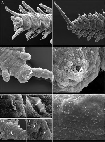
Description Fig. 10 Scanning electron microscopy images of Ramisyllis kingghidorahi n. sp., posterior-most regions and epithelium details. A–D Posterior ends. Arrow in C and D points to heavily ciliated anus. E– G Minute crests on the dorsal surface of midbody segments. Arrows
point to crests laterally located on the dorsal surface. H Dorsal surface of posterior segments. I Clumps of cilia on dorsal surface of proventricular segments. Arrows pointing to pores in H. Scale bars: 100 μm A, B, I, 50 um C, G, 5 μm D, E,4 μm F, and 3 μm H
Source: Aguado, M. T.; Ponz-Segrelles, G.; Glasby, C. J.; Ribeiro, R. P.; Nakamura, M.; Oguchi, K.; Omori, A.; Kohtsuka, H.; Fischer, C.; Ise, Y.; Jimi, N.; Miura, T. (2022). Ramisyllis kingghidorahi n. sp., a new branching annelid from Japan. Organisms Diversity & Evolution. 22(2): 377-405., available online at https://link.springer.com/article/10.1007%2Fs13127-021-00538-4
Copyright/ownership information for image: Organisms Diversity & Evolution Journal
·
Click here to return to the thumbnails overview

