Integrated Marine Information System (IMIS)
Persons | Institutes | Publications | Projects | DatasetsSEM images of spicules
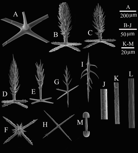
Description A, the centre part of choanosomal pentactin; B–C, atrial pinular pentactin; D, dermal pinular pentactin; E–F, choanosomal pinular pentactin; G, pinular hexactin; H, stauractin; I, spiny microdiactin; J, smooth middle shaft of marginalia; K, details of the middle shaft of mesouncinate; L, details of the middle shaft of macrouncinate; M, micramphidisc.
JPG file - 1.29 MB - 2 421 x 2 689 pixels
Extra information
FileName: 149668.jpg
FileDateTime: 1635597088
FileSize: 1289443
FileType: 2
MimeType: image/jpeg
SectionsFound: ANY_TAG, IFD0, THUMBNAIL, EXIF
COMPUTED.html: width="2421" height="2689"
COMPUTED.Height: 2689
COMPUTED.Width: 2421
COMPUTED.IsColor: 1
COMPUTED.ByteOrderMotorola: 1
COMPUTED.Thumbnail.FileType: 2
COMPUTED.Thumbnail.MimeType: image/jpeg
Orientation: 1
XResolution: 3000000/10000
YResolution: 3000000/10000
ResolutionUnit: 2
Software: Adobe Photoshop CS6 (Windows)
DateTime: 2015:11:04 21:45:43
Exif_IFD_Pointer: 164
ColorSpace: 1
ExifImageWidth: 2421
ExifImageLength: 2689
added on 2021-10-30540 views
FileName: 149668.jpg
FileDateTime: 1635597088
FileSize: 1289443
FileType: 2
MimeType: image/jpeg
SectionsFound: ANY_TAG, IFD0, THUMBNAIL, EXIF
COMPUTED.html: width="2421" height="2689"
COMPUTED.Height: 2689
COMPUTED.Width: 2421
COMPUTED.IsColor: 1
COMPUTED.ByteOrderMotorola: 1
COMPUTED.Thumbnail.FileType: 2
COMPUTED.Thumbnail.MimeType: image/jpeg
Orientation: 1
XResolution: 3000000/10000
YResolution: 3000000/10000
ResolutionUnit: 2
Software: Adobe Photoshop CS6 (Windows)
DateTime: 2015:11:04 21:45:43
Exif_IFD_Pointer: 164
ColorSpace: 1
ExifImageWidth: 2421
ExifImageLength: 2689
Click here to return to the thumbnails overview
