Integrated Marine Information System (IMIS)
Persons | Institutes | Publications | Projects | DatasetsCaldithrix abyssi strain LF13T
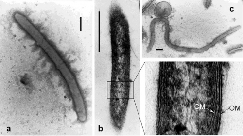
Description (a) Electron micrograph of a negatively stained cell of strain LF13T. Bar, 0.5 mm.(b) Electron micrograph of thinsection of a cell of strain LF13T, exhibiting cell-wall structure. CM, cytoplasmic membrane; OM, outer membrane. Bar, 0.5 mm.
(c) Electron micrograph of thin-section of a cell of strain LF13T, exhibiting formation of a spherical body. Bar, 0.5 mm.
Source: https://doi.org/10.1099/ijs.0.02390-0
JPG file - 120.43 kB - 886 x 499 pixels added on 2018-06-291 424 views
Click here to return to the thumbnails overview

