WoRMS Photogallery

Neoechiniscoides aski Møbjerg, Jørgensen & Kristensen, 2020
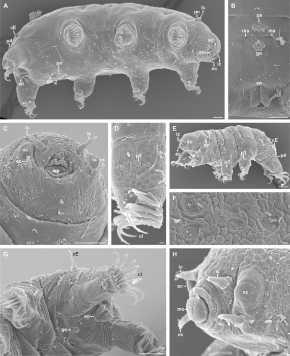
Description Figure 3. Scanning electron microscopy of Neoechiniscoides aski. A–C, female. A, habitus of mature female. Scale bar: 10 μm. B, close-up of gonopore with associated cuticular plate and anus. Note muscle attachment sites located lateral to the female gonopore. Scale bar: 10 μm. C, close-up of head region. Note the cuticular sculpture of the head. Scale bar: 10 μm. D–H, male. D, second leg, with dome-shaped sense organ (p2). Scale bar: 1 μm. E, habitus of adult male. Scale bar: 10 μm. F, prominent dorsal cuticular attachment sites of the caudal segment. Scale bar: 1 μm. G, posterioventral part of a male, showing the fourth leg pair, anal system and gonopore. Anterior is to the left. Scale bar: 10 μm. H, close-up of head region of male. Note the relatively large secondary clavae. Scale bar: 1 μm. Abbreviations: an, anus; cE, cirrus E; cl, claw; ec, externalLink to publication: https://www.marinespecies.org/aphia.php?p=sourcedetails&id=391897
PNG file - 505.48 kB - 595 x 731 pixels added on 2021-03-151 874 viewsWoRMS taxaMicroscope Neoechiniscoides aski Møbjerg, Jørgensen & Kristensen, 2020checked Kristensen, Reinhardt Møbjerg 2021-03-19
This work is licensed under a Creative Commons Attribution-NonCommercial-ShareAlike 4.0 International License
Click here to return to the thumbnails overview
 Comment (0)
 Click here to add a comment.
* indicates a required field.
Disclaimer: WoRMS does not exercise any editorial control over the information displayed here. However, if you come across any misidentifications, spelling mistakes or low quality pictures, your comments would be very much appreciated. You can reach us by emailing info@marinespecies.org or adding a comment, we will correct the information or remove the image from the website when necessary or in case of doubt.