WoRMS Photogallery

Cladocarpus unilateralis
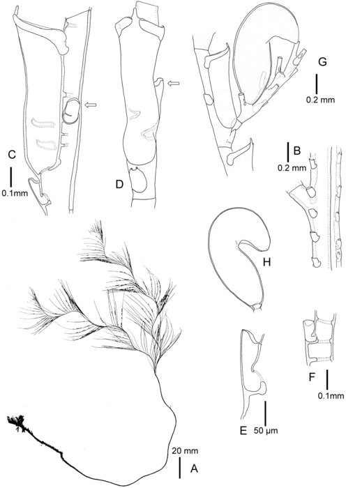
Description Holotype. (A) Colony silhouette. (B) Part of polysiphonic stem with main tube in front (with apophysis) and an auxiliary tube in rear. (C) Hydrothecate segment in lateral view, note lateral median nematotheca (arrow). (D) Hydrothecate segment in frontal view, note lateral median nematotheca (arrow); same scale as C. (E) Median inferior nematotheca of hydrothecate segment. (F) Unilateral nematotheca of hydrothecate segment seen from rear side. (G) Hydrothecate segment with phylactocarp bearing a gonotheca. (H) Gonotheca in side view; same scale as G.
Source: Schuchert P. 2015. On some hydroids (Cnidaria, Hydrozoa) from the Okinawa Islands, Japan. Revue Suisse de Zoologie 122: 325-370. www.doi.org/10.5281/zenodo.30004
PNG file - 227.18 kB - 5 020 x 7 087 pixels added on 2025-02-11123 viewsWoRMS taxaScan of drawing Cladocarpus unilateralis Schuchert, 2015checked Schuchert, Peter 2025-02-11 Download full size

This work is licensed under a Creative Commons Attribution-NonCommercial-ShareAlike 4.0 International License
Click here to return to the thumbnails overview
 Comment (0)
 Click here to add a comment.
* indicates a required field.
Disclaimer: WoRMS does not exercise any editorial control over the information displayed here. However, if you come across any misidentifications, spelling mistakes or low quality pictures, your comments would be very much appreciated. You can reach us by emailing info@marinespecies.org or adding a comment, we will correct the information or remove the image from the website when necessary or in case of doubt.