WoRMS Photogallery

Tripaphylus squidwardi Boxshall, Barton, Kirke, Zhu & Johnson, 2022
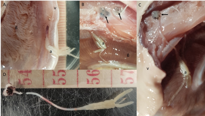
Description Fig. 1 Female specimen of Tripaphylus squidwardi collected from Carcharhinus coatesi. A Female embedded into the ventral gill flap with the abdomen visible in region of the gill. Tape measure indicates millimetres. B Ventral skin removed from the throat region of the shark, exposing the head (arrow) of two specimens of Tripaphylus squidwardi. C A female T. squidwardi fully dissected from the ventral throat region of the shark in situ. D A female T. squidwardi removed from the shark. Tape measure indicates centimetres. a, abdomen of copepod; g, gill of shark; v, ventral surface of shark
Source: Barton, D.P., A. Kirke, G. Johnson & G. Boxshall. (2022). Pathology associated with Tripaphylus Richiardi, 1878 infection (Copepoda: Sphyriidae) in wild-caught Australian blackspot sharks, Carcharhinus coatesi (Whitley, 1939), off northern Australia. Parasitology Research. 121(11):3337-3340. Sep 2022., available online at https://doi.org/10.1007/s00436-022-07662-w
·
This work is licensed under a Creative Commons Attribution-NonCommercial-ShareAlike 4.0 International License
Click here to return to the thumbnails overview
 Comment (0)
 Click here to add a comment.
* indicates a required field.
Disclaimer: WoRMS does not exercise any editorial control over the information displayed here. However, if you come across any misidentifications, spelling mistakes or low quality pictures, your comments would be very much appreciated. You can reach us by emailing info@marinespecies.org or adding a comment, we will correct the information or remove the image from the website when necessary or in case of doubt.