WoRMS Photogallery
Phaenocora evelinae
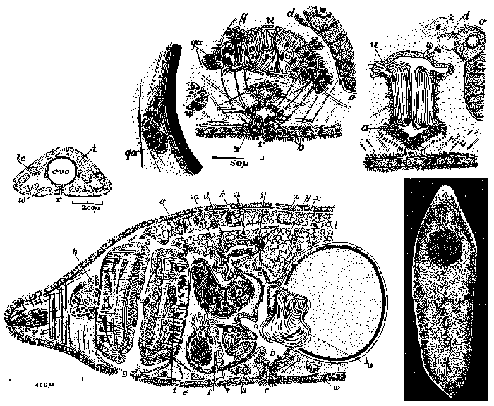
Description Fig. 84 (below). Sagittal section of the anterior region. Combination of a phase with an egg in the uterus with another, after oviposition, with the pyriform appendix contracted. a, atrio superior. b, atrio inferior. c, canal genital feminino. d, oviduto. e, duto eferente. f, diafragma. h, cerebro. i, intestino, k, clavas granulosas de Minot. m, bursa. n, duto bursa-intestinal. o, ovario. p, poro bursa-intestinal. r, gonoporo. s, glandulas da secrecao granulosa. t, reservatorio da secrecao granulosa. u, apendices piriformes. v, vesicula seminal. w, vitelario. x, diverticulo do canal genital feminino. y, esfincter ectal do canal genital feminino. z, glandula lobulada, uninucleada.
Fig. 88 (small drawing at the left). Transverse section of the anterior third.
Fig. 89-90 (two upper right drawings). Two transverse sections situated close together, of the same series. Two pyriform appendages and atrium superior. GIF file - 81.81 kB - 1 205 x 993 pixels added on 2017-04-0754 viewsWoRMS taxaScan of photo Phaenocora evelinae Marcus, 1946checked Tyler, Seth 2017-04-07
Click here to return to the thumbnails overview
 Comment (0)
 Click here to add a comment.
* indicates a required field.
Disclaimer: WoRMS does not exercise any editorial control over the information displayed here. However, if you come across any misidentifications, spelling mistakes or low quality pictures, your comments would be very much appreciated. You can reach us by emailing info@marinespecies.org or adding a comment, we will correct the information or remove the image from the website when necessary or in case of doubt.
