WoRMS Photogallery
P. busheki
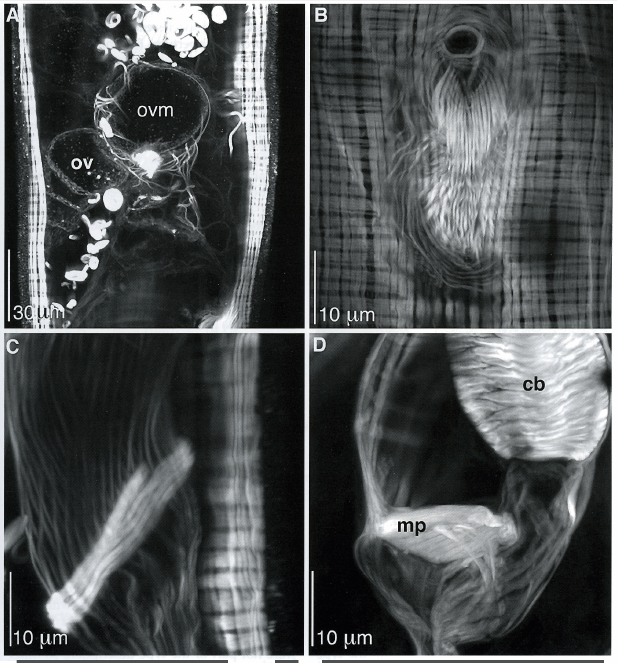
Description Z-projections of reproductive anatomy of P. busheki Ax. A. Anterior end of oviduct where it 'opens' to the developing ova. 0.1?m x 100 optical sections. B. Receptaculum seminis, showing female pore and fan-like array of longitudinal muscles. 0.05?m x 60 optical sections. C. Sphincter muscles at the base of the uterus (top of female genital canal). 0.05?m x 179 optical sections. D. Unusual plate-like structure of stacked muscle cells within the male copulatory apparatus. 0.05?m x 107 optical sections
PNG file - 705.50 kB - 617 x 663 pixels
added on 2017-04-07657 viewsWoRMS taxaScan of photo Prognathorhynchus busheki Ax, 1997checked Tyler, Seth 2017-04-07
Click here to return to the thumbnails overview
 Comment (0)
 Click here to add a comment.
* indicates a required field.
Disclaimer: WoRMS does not exercise any editorial control over the information displayed here. However, if you come across any misidentifications, spelling mistakes or low quality pictures, your comments would be very much appreciated. You can reach us by emailing info@marinespecies.org or adding a comment, we will correct the information or remove the image from the website when necessary or in case of doubt.
